Glaucoma is a disease process that damages the optic nerve of the eye.

The optic nerve is a cable of about a million nerve fibers acting like electrical wires that transmit the visual information received by the eye to the brain for processing. Glaucoma is a disease process that involves loss of these nerve fibers and is the second leading cause of blindness worldwide.  It is usually painless, slow, and permanent giving it the moniker the “silent thief of sight.” There are often no symptoms until there is significant, permanent vision loss. Fortunately, through routine dilated eye exams and intra-ocular pressure checks, we can diagnose this condition, and, if treated early, we can prevent blindness.

Types of Glaucoma

  • Primary Open Angle Glaucoma (POAG): This is the most common form of glaucoma. It is identified by increase IOP and a progressive change in the optic nerve while the drainage system structure is normal and the eye is healthy with no underlying disease. POAG occurs slowly over many years and has no symptoms.
  • Closed-Angle Glaucoma: This type of glaucoma is much less common and can be sudden and painful. In this type of glaucoma, the iris blocks the drainage structure within the eye preventing aqueous outflow leading to a sudden and significant increase in IOP. This can be a glaucoma emergency and you need to call right away if you notice symptoms including sudden blurry vision with halos around lights, severe eye pain, and halos around lights. You may even develop headaches and vomiting due to the severe pain.
  • Neovascular Glaucoma (NVG): This is a form of secondary glaucoma as it is due to another underlying condition such as diabetes, central retinal vein occlusion, or carotid artery obstructive disease. With such diseases, the small blood vessels within the eye degenerate and close off and the eye becomes starved for oxygen and nutrients normally provided by normal blood flow. In response, the eye creates new blood vessels that unfortunately cause more harm than good as they grow in a uncontrolled fashion. These blood vessels grow over the drainage structure inside the eye blocking the aqueous drainage much like getting hair blocking a shower drain leading to sudden increase in eye pressure.
  • Juvenile Glaucoma/Congenital Glaucoma: This is a rare condition that occurs in children typically less than 1 year old.

Causes 

While most types of glaucoma deal with increase intraocular pressure (IOP), not all glaucoma patients have high IOP. The optic nerve is incredibly vulnerable to damage from elevated eye pressure. Over time, this high IOP can cause a loss of the individual nerve fibers that make up the optic nerve leading to a loss of vision and ultimately, blindness.

Clear fluid (aqueous) circulates within the front part of the eye nourishing the cells and structures of the eye. This fluid is constantly produced at the same rate that it drains from the eye thus maintaining a constant pressure. The fluid movement within the eye is much like a faucet. The aqueous is produced by an organ behind the iris, flows through the pupil, and exits at a drainage structure located in front of the iris. This fluid is not part of the tears on the outer part of the eye and has no relation to tearing or dry eyes.

The IOP can increase with either an overproduction of the aqueous or a blockage in the drainage of the aqueous from the eye. When the pressure is elevated, irreversible damage occurs to the optic nerve. The primary goal of any glaucoma treatment is to lower the pressure to a safe level for the optic nerve.

Diagnosis

Through a routine dilated exam with intraocular pressure check and evaluation of the optic nerve, glaucoma can be diagnosed; however, the diagnosis is not always an easy one to make. Because the disease progresses so slowly most of the time, it may take several exams over time to look for a change in the optic nerve as well as multiple IOP measurements to be confident in the diagnosis. The IOP fluctuates throughout the day and high IOP can easily be missed on just one examination. Several other risk factors are considered with making the diagnosis of glaucoma as well.

Risk Factors:

  • Increasing age, especially after age 60
  • Family history of glaucoma
  • Elevated Eye Pressure
  • African or Spanish-American descent
  • Near-sightedness and Far-sightedness
  • Previous Eye Injuries
  • Thinner central corneal thickness

Advanced testing can also be performed to detect early visual field defects and damage to the optic nerve fibers to aid in the earliest possible detection. Putting all of the risk factors, signs, and symptoms together, a diagnosis of glaucoma can be made. If you have a few risk factors, you may be diagnosed as a glaucoma suspect and require closer monitoring as you may be at a higher chance of developing the disease.  Because of the lack of symptoms, annual eye examinations are very important, especially after age 60.

Treatment Options

Damage caused by glaucoma is irreversible; thus, early detection and treatment are essential. Fortunately, there are many treatment options for today’s glaucoma patient.

  • Eye drops
  • Oral medications
  • Laser treatments
  • Surgery including Micro-invasive glaucoma surgery (MIGS), trabeculectomy, goniotomy, tube shunt placement and more options are being developed.  Even cataract surgery can be beneficial to lower eye pressure.

Eye Drops

Most patients with glaucoma are easily managed with eye drop therapy. It may take only one type of drop, but more advance glaucoma may require multiple drops. These drops work to decrease production of aqueous and/or increase the drainage of aqueous out of the eye.

Possible side effect of glaucoma eye drops

  • Stinging or burning
  • Red eyes or redness of the skin around the eyes
  • Change in heart rate
  • Changes in breathing especially in those already compromised with COPD, asthma, emphysema
  • Dry mouth
  • Blurred vision
  • Change in eye color and skin color around the eye
  • Longer eyelash growth

Laser Surgery

Laser surgery treatments for glaucoma are done for two different reasons for 2 different types of glaucoma.

  1. POAG: The laser treatment is directed toward the drainage structure of the eye to open the drainage channels allowing for increased outflow to reduce the IOP. This treatment can be performed multiple times.
  2. Closed-angle glaucoma: The laser treatment is directed towards creating an opening or hole in the iris providing a secondary path for fluid to flow to the drainage structure.

Surgery in the Operating Room

In the past, surgical glaucoma treatment was limited to invasive and somewhat risky procedures that were limited to severe glaucoma patients only. Over the past 20 years, a myriad of different, less invasive, and safer surgical options have become available. Because of this, we can now offer glaucoma surgery for those with mild to moderate glaucoma.

Minimally invasive glaucoma surgery (MIGS) is a group of different types of surgical treatments for glaucoma that are much less invasive and have a significantly improved safety profile compared to many other glaucoma procedures. With all glaucoma treatments including surgery, the goal is to reduce the intra-ocular pressure (IOP) that is putting too much stress on the optic nerve. Previously, surgical intervention was reserved for those glaucoma patients with severe glaucoma or who were on maximal medical therapy and still having progressive vision loss.

With the advent of MIGS procedures, very safe and effect surgical treatments are now being used much earlier in the treatment of glaucoma allowing for better IOP control. One such procedure involves the iStent Inject W.

iStent Inject® W

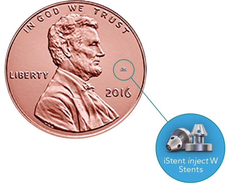
The iStent Inject® W is quite possibly one of the smallest medical devices everimplanted into humans. It functions to restore the eye’s natural ability to drain fluid from the front part of the eye helping to reduce the intra-ocular pressure providing improved glaucoma outcomes.

Who’s a Candidate:

  • For those with mild to moderate glaucoma and in need of cataract surgery, the iStent Inject® W may be a great option for you as this implant can be placed safely and easily at the time of cataract surgery providing you with additional benefit to your time in the operating room.

Benefits:

  • iStent Inject® W, once implanted, works continuously providing reduced IOP 24 hours a day.
  • For those on glaucoma eye drops, implanting the iStent Inject® W may help reduce the number of glaucoma medications needed.

How iStent Inject® W Works

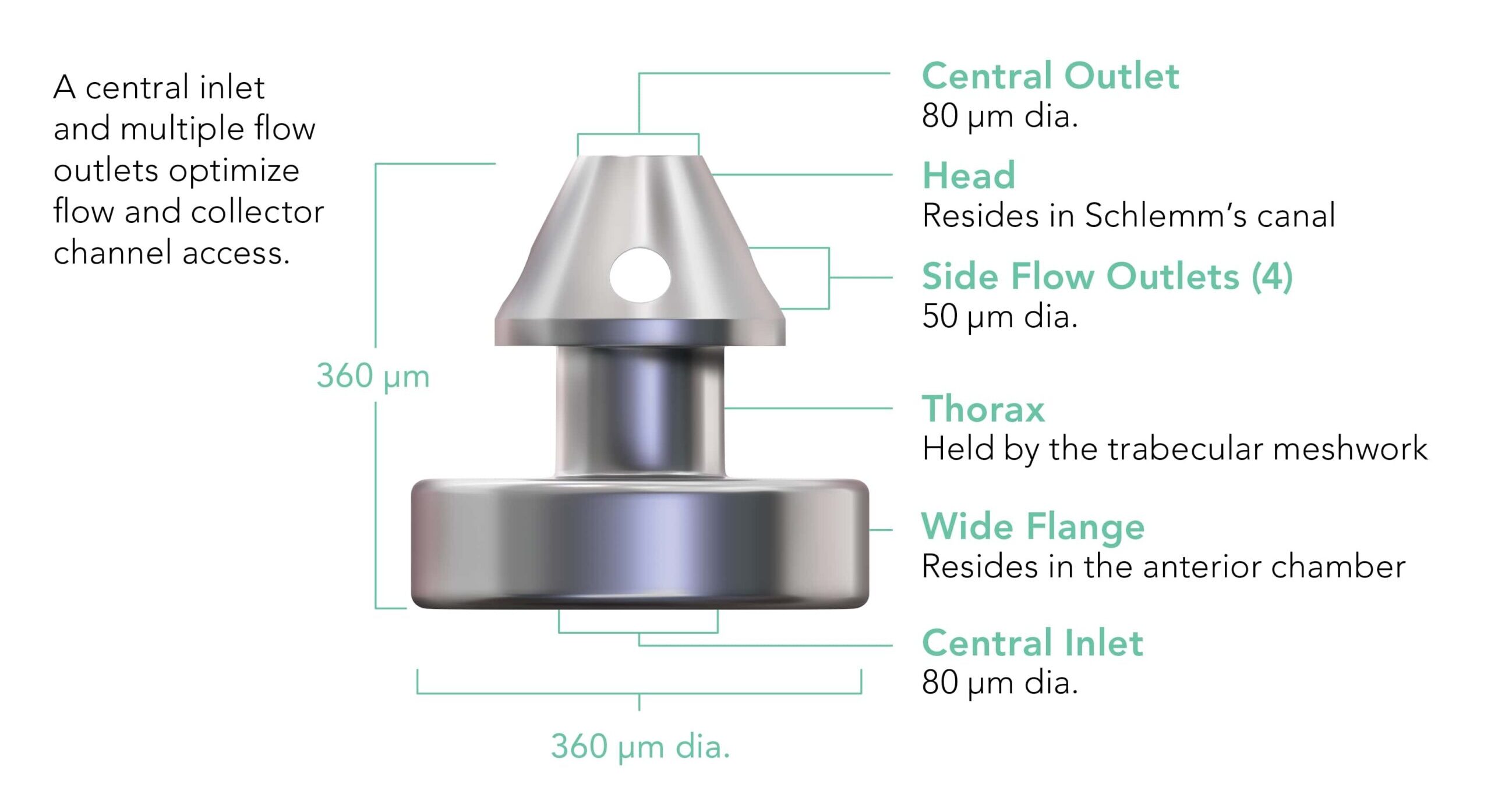
  • The iStent Inject® W includes two surgical-grade titanium stents that are less than 0.5mm in size.
  • Once your cataract surgery is completed, two iStent Inject® W implants are placed in the poorly functioning drainage apparatus in your eye called the trabecular meshwork. The stents provide 2 bypasses, or openings, between the front part of the eye and its natural drainage pathway increasing the flow of fluid.

Risks of the iStent Inject® W

  • Stent blockage preventing proper functioning and reduced benefit
  • Intraocular inflammation risk slightly higher than with cataract surgery alone
  • Secondary surgical intervention
istent
istent

Accessibility Toolbar